3D MODELING SHOWS RELOCALIZATION OF HETEROCHROMATIC REPAIR FOCI


A Drosophila cell has been irradiated to induce DSB formation, and monitored by time-lapse microscopy. 3D modeling of heterochromatin and repair proteins has been obtained with Imaris, and it shows relocalization of repair centers to the euchromatic space (arrows). Numbers = minutes after irradiation. Scale bar = 1um
LATE REPAIR STEPS OCCUR OUTSIDE HETEROCHROMATIN
A Drosophila cell shows late repair steps (Rad51 foci) occurring outside the heterochromatin domain. Top left: detail of one Rad51 focus highlights its dynamics in the euchromatic space. The first image (no foci) is taken before DSB induction.

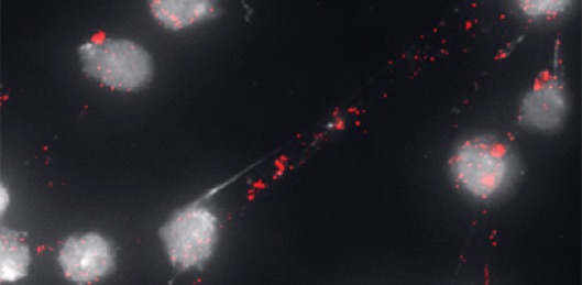
SMC5/6 PREVENTS ABNORMAL RECOMBINATION IN HETEROCHROMATIN
The Smc5/6 complex is required to control the progression of HR in heterochromatin. This image shows Drosophila cells stained for DNA and heterochromatin. Smc5/6 loss results in aberrant HR (heterochromatic DNA filaments connecting dividing cells).

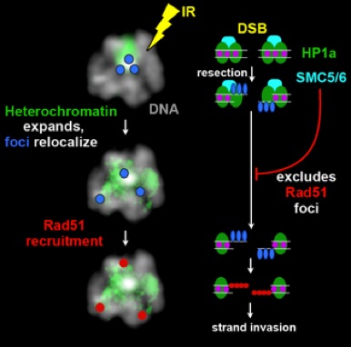
MODEL OF SPATIAL AND TEMPORAL CONTROL OF REPAIR IN HETEROCHROMATIN
The initial processing of heterochromatic DSBs occurs quickly in heterochromatin. Later repair steps, including Rad51 recruitment and strand invasion, are suppressed by the Smc5/6 complex. Next, heterochromatin expansion promotes relocalization of repair sites.
The euchromatic environment allows Rad51 loading and completion of HR repair.
We hypothesize that this mechanism prevents ectopic recombination by isolating the damaged repeated DNAs from the rest of heterochromatin during repair